Author: DVM Adina Argăseală
If in the previous article we discussed the importance of an ophthalmological examination in pets, in what follows describing in detail what this consultation entails, what work and their role in the objective evaluation of the visual apparatus are.
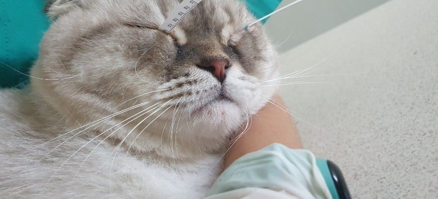
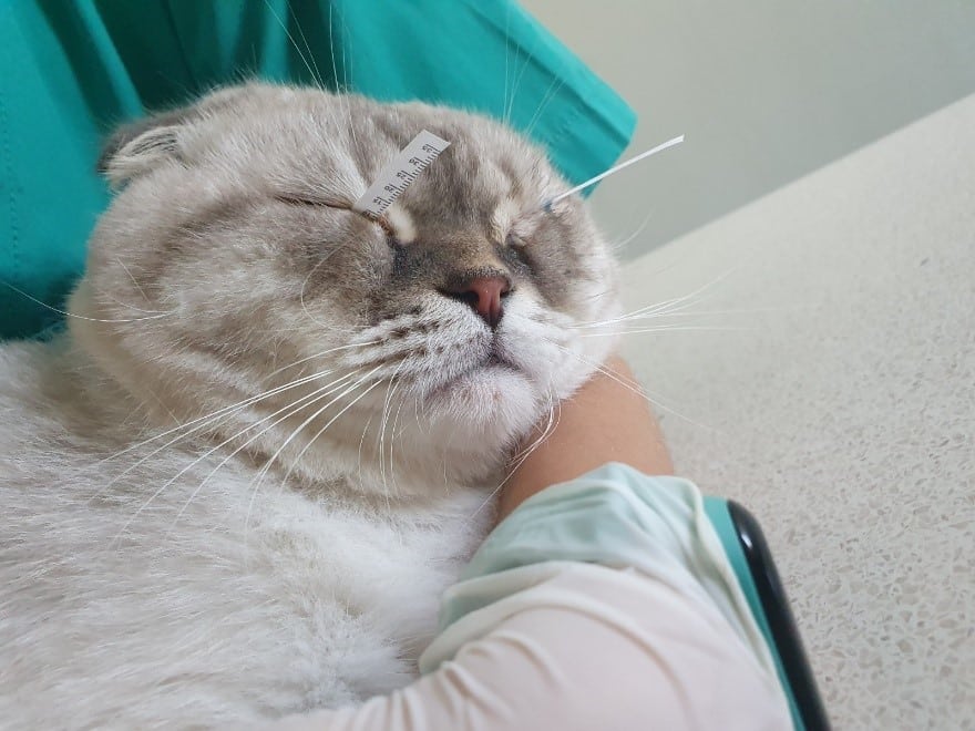
Inside the veterinarian for pets, the ophthalmological examination is carried out in a dark room, using specialized equipment. All the work in the ophthalmological exam is painless, does not require sedation for cooperative patients. A usual examination of a new patient takes about 30-40 minutes, the duration extends if further investigations are needed.
The complete ophthalmological exam provides several phases, the first of them is a detailed anamnesis in which we want to collect all the information on the ophthalmological condition, the patient’s general conditions and other conditions diagnosed in the past.
View Test It consists in pursuing the response to the threat, the test with cotton pieces, where you can see how the patient moves in a foreign environment, if it affects the obstacles, if it climbs and descends the stairs, both on the light and in the dark.
For Examination of the previous segment Biomicroscope is used (crack lamp), which allows the detailed evaluation of the eyelids, conjunctiva, cornea, aqueous humor, Iris and crystalline.
The evaluation of the bottom of the eyes is composed of Direct ophthalmoscopy Using the panoptic ophthalmoscope. Some patients may require dilation of pupils, it is made with a half medium -aciatea which with atropine. Sometimes the drops must be instilled several times, until the pupil is completely expanded, the time can vary depending on the patient.
Under the action of light, the student should reduce (Reflection of the pupillary photomotor). If this does not happen, it can indicate a certain problem, depending on the light used, blue or red. The CPLR tester uses the light of a certain intensity and wavelength, stimulating in particular the chapter and lymph nodes, provides information on the state of the retina and optical nerve. Reflexes that involve some cranial nerves are also verified, therefore the ophthalmological examination can also be useful in the diagnosis of neurological conditions.
TometryThe measurement of intraocular voltage is particularly important, being useful in the detection of glaucoma (when the pressure values have increased) or uveite (when the values are low).
The quantitative or qualitative deficit of tears (dry eye syndrome) is very common and can predispose to corneal disorders, such as keratitis, corneal ulcers. The film Lacrima is evaluated from a quantitative point of view using The skiers’ test and qualitatively using the method Running time of the tear film (asking for fluorescent and fluorescent light of the biomicroscope).
Installation of a drop of fluorescein Highlights the presence of possible corneal injuries. All with the help of fluorescein is performed e Jones testthus checking the permeability of the teasal-nasal channels. In the case of patients with epifora (excessive tears), the tearcrous points can be controlled and permeable in local anesthesia, if the patient allows it.
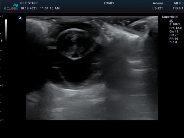
Depending on the results of the tests described above, it is possible to recommend further tests that can be performed within the hospital at the time of consultation or later. For example, if the direct evaluation of the eye or orbit is not possible, it is used Eyeball. The procedure is painless, most patients collaborate. This is thus achieved important details relating to the crystalline, vitreous, retina body, in the presence of tumor formations, orbital abscesses, etc.
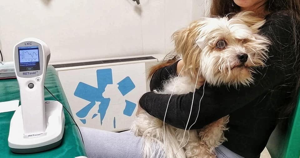
Electroretinography represents the evaluation of the electric function of the retina. It is performed mandatory before the cataract operation, in the case of patients with suspicion of retinal atrophy (when they may have nocturnal or day vision deficits) and in the event of sudden blindness in which you want to differentiate between retinal blindness or a secondary blindness to optic blindness in most of the cooperative patients, sedation is not required.
Some ophthalmological conditions can be a symptom in systemic conditions, such as infectious diseases, hypertension, diabetes, neoplasm. In this case they are requested Further investigationsAs blood tests, bacteriological examination, cardiological examination, abdominal ultrasound, radiographic examination, imaging (CT, MRI).
At the end of the exam, observation sheet which is delivered to the owner includes: the results of each phase of the ophthalmological examination, presumptive or certainty, information on the treatment and their administration, an estimated cost relating to the investigations/procedures recommended. The images with eye injuries and the lower part of the eye are connected to the digital sheet and are accessible by the owner at any time, in order to follow the dynamic evolution.
In conclusion, a specialized ophthalmological examination includes a series of painless maneuvers, most of the time it can be done without sedation and after an overview of the entire visual system and some neurological pathologies (if applicable) has been obtained. The early diagnosis of pathologies at this level and the establishment of adequate treatment will improve the quality of life of our quadrupeds and avoid the appearance of irreparable changes.
latest posts published
The health of the joints in dogs – more important than you would have thought!

Everything you need to know about Dog & Dog – Dry Dog Food

Obesity in dogs and what should be done?
The dog needs to have a happy, balanced and healthy pet

How do you choose and how do you use cat sand?

How do we help pets during winter holidays, full of strong noises and noises

The benefits of fish in the diet of our empty spaces

The joy of having a cat

Golden Retriever, an energetic and favorite breed of children