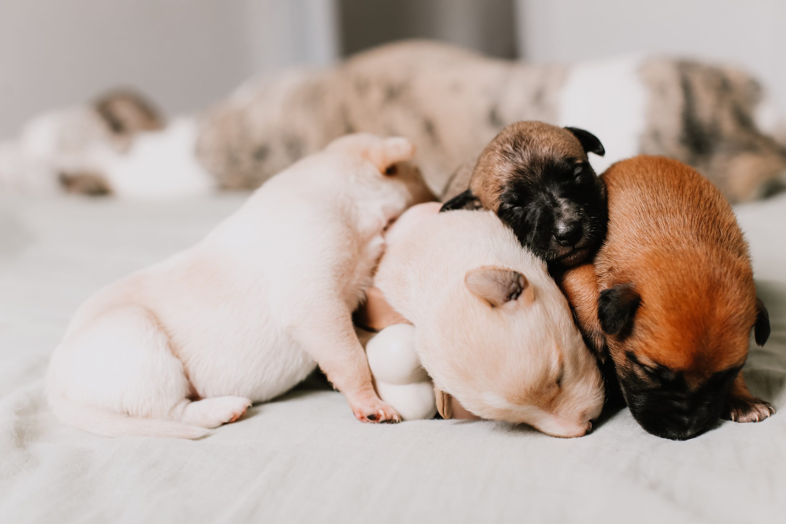
The eye check-up has become more frequent for pets, as they are important parts of the family. Vision helps them navigate their surroundings and carry out daily tasks effectively.
AND Eye check-up It should be done every year for animals without any specific signs of eye issues and at least every 6 months for older pets. A basic eye exam is also suggested during the first visit of the pet to the vet, after being separated from the litter. Some conditions can be passed down from parents or may develop shortly after birth, needing a diagnosis and treatment as soon as possible.
The importance of the ophthalmological examination in pets
The general vet should look at the matching between the two eyes, the ability to track nearby objects, and the presence of the eyelid response, meaning the closing of the eyelids when they touch them.
It is also assessed if the patient has a certain degree of entroponio (curbing the eyelids inwards) or ectropion (restoring the eyelids outside, with highlighting the conjunctiva at a higher level). You can appreciate the pupil response to a bright stimulus and a butt exam per eye can be done using a direct ophthalmoscope. If the general doctor observes some changes and considers it necessary, it can refer to a doctor with more experience in veterinary ophthalmology.
read more Period of heat in cats: everything you need to know about the reproductive cycle.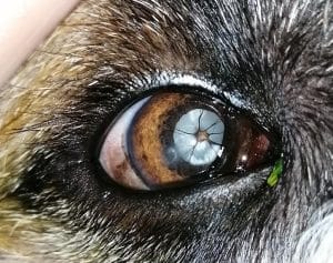
There are some breeds of dogs and cats inclined to some specific ophthalmological conditions, for them the visit to the recommended obessment when the first signs of eye discomfort.
Entropion
This condition usually occurs in brachycephalic breeds, in both dogs and cats, but it can also appear in other types. In severe situations, which might happen in young animals, the eyes can become almost fully closed, leading to corneal ulcers. Surgery is often the only treatment option. Sometimes temporary measures like using tacks, surgical clips, or hyaluronic acid injections in the eyelids can be used until the patient can undergo surgery.
Some objective issues (Cataract, lens dislocation) or glaucoma can be present from birth and may show up early in an animal’s life. If not identified and treated promptly, these problems can result in serious complications, potentially causing blindness or even loss of the eye.
Dogs with glaucoma, especially those breeds like Jack Russell Terrier, Cocker Spaniel, Husky, Akita, Samoyed, and Basset Hound, should be examined using a specific method called Gonoscopy. Using a special lens, the angle between the cornea and iris can be inspected to see if there are any changes in the normal eye structure. If any abnormalities are found, it is advisable to use preventive antiglaucomatous medications.
read more How do you choose the veterinary clinic suitable for your pet?
Additionally, issues at the level of the retina, the part of the eye that captures visual information and has a genetic factor, can appear in some breeds. These include Progressive retinal atrophy, common in Labrador/Golden Retriever breeds, Cocker Spaniel, and German Shorthaired Pointer, as well as retinal dysplasia and degeneration of the retina.
A suspected diagnosis can be made from the medical history and eye examination. For a definitive diagnosis of these conditions, a specialized test called electroretinography is necessary, which assesses the electrical activity in the retina, similar to the electrocardiogram used for heart checks.
When does the ophthalmological examination become an emergency?
- Trauma In the cranial region involving the eyebrows of the eye bulbs:
- Road accidents, bites caused by other dogs, which can lead to the dislocation or invulsion of the eyeball (its exit). In this situation, a medical consultation for the patient is recommended, in order to stabilize the patient and decide if the eye can be saved by a repositioning surgery
- Cut/spread horny wounds, produced by sharpened foreign bodies or by an cat’s claw
- Clinical signs can manifest themselves from intense pain, dark eyes, excessive tears, small pupils, presence of blood inside the eyeball, which can sometimes be outsourced. The asset examination as quickly as possible can make a diagnosis of certainty, after which the therapeutic behavior will be established. Most of the time it is also recommended to perform an ocular ultrasound, to see if the crystalline has been influenced accordingly, post -traumatic cataract can occur or if the detachment of the retina has occurred. The treatment is often surgical, with the suture of the cornea and the application of a biomaterial that protects the surface of the eyeball and haste the healing.
- Acute glaucoma: The owner can see a sudden opacification on the surface of the eyeball (corneal edema), the pain manifested by tear, keeping the eye dark (blefarospas), sensitivity to light (photophobia). Tonometry is the only specific method capable of determining the value of intraocular tension, after which it is possible to establish the diagnosis of glaucoma and it is possible to decide adequate treatment. After receiving this diagnosis, the riflete will be carried out for short periods of time to verify the efficiency of the treatment established.
- Diabetic cataract: Patients with diagnosis of diabetes, in particular dogs, can develop cataracts at a range of several months. The owners observe the reduction of vision, with the stroke of obstacles, the opacification in the objective. Sometimes eye signs are the first ones who appear in case of diabetes. Secondary diabetic cataracts, wealthy raisins, a rather serious intraocular inflammation that requires early treatment to avoid subsequent complications and therefore increase the patient’s possibilities to become a candidate for cataract surgery can occur.
- Sudden blindness: Usually occurs due to retina disorders or level of the optical nerve. At the level of retina it can concern the degeneration of the acute retina, which leads to blindness in a short period of time. Most of the time it affects dogs, no changes are observed in the ophthalmological examination, only after the electrinography has been able to establish the diagnosis. The sudden loss of vision can be produced after the grip of the retina, which usually occurs after a serious or secondary ocular trauma with serious intraocular changes.
- In addition, a more common aspect in geriatric cats with heart or kidney disease can occur consecutively to systemic hypertension. With a correct diagnosis and an adequate treatment of systemic disease, there are situations in which the retina can be recalled, the patient is able to recover his partial vision.
- Optical neurita It is a serious inflammation of the optical nerve, which makes the secondary vision lose. The suspicion of this diagnosis can do it after the ophthalmological examination, but further investigations are recommended for a diagnosis of certainty: electrinography, cranial magnetic resonance imaging.
The ophthalmological examination can be done in other species of animals, including exotic ones, many of whom are considered family members. The tests that can be performed are similar to those of the dog and the cat, others adapted according to the patient.
read more
 The importance of blood tests in pets
latest posts published
The health of the joints in dogs – more important than you would have thought!

Everything you need to know about Dog & Dog – Dry Dog Food
Obesity in dogs and what should be done?
The dog needs to have a happy, balanced and healthy pet
How do you choose and how do you use cat sand?
How do we help pets during winter holidays, full of strong noises and noises
The benefits of fish in the diet of our empty spaces
The joy of having a cat
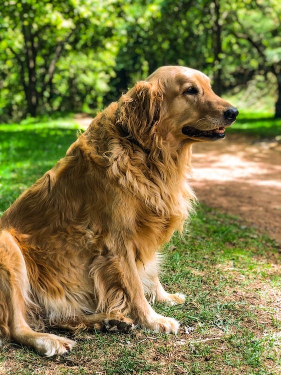
Golden Retriever, an energetic and favorite breed of children