Author: Dr. Alexis Gîrloanțț
Regardless of the color of the animal’s fur, a specific pigmentation of the hair in the periocular area is very common, caused by the secretion of tears (tears).
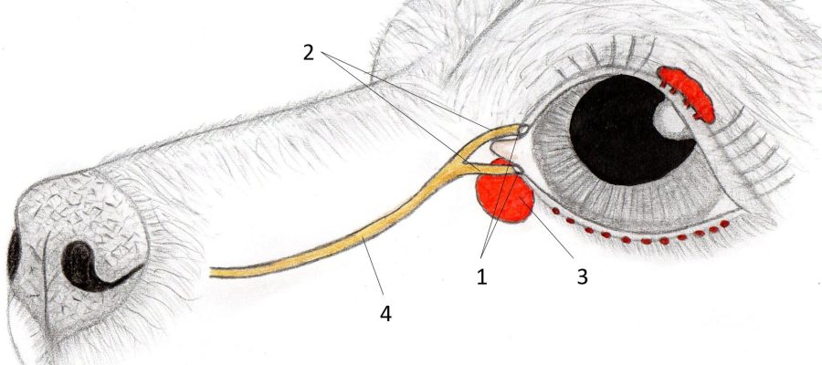
The lacrimal apparatus is composed of: The main tear gland (located in the upper eyelid region), The attached gland of the third eyelid (Located at the level of the third eyelid), Meibomius glands (located on the free edge of the eyelids) e Pious of tears. The secretions of these glands form the tear film of the devolve, which has the main role of corneal protection.
The reddish/brown color of the fur around the eyes may be due to losses a greater amount of tears in the periocular area, this clinical sign is called tearing. Although the tears are transparent, the hair on which they restore for a long time change their color due to a substance called lactoferin What is naturally in the composition of the tear secretion and easily «spots» the animal fur, especially if it is white. It is also suspected that bacteria on the skin also participate in this color.
The stagnation of tears in the adjustment area determines the creation of a humid environment that leads to the multiplication of bacteria/mushrooms and implicitly can complicate the epiphor with the appearance Blefaritelor (inflammation of the eyelids) or dermatitis.
What does the epiphara appearance determine?
1.

The tear system consists of 2 tears, 2 tears and a nasolacrimal channels, which aim to transport tears from the conjunctival bag to the nasal cavity, its obstacles that translate into tears on the periocular surface.
2. Irritive factors
The most common irritating factor is represented by hair which, by contact with cornea and conjunctiva, determine an excessive secretion of tears. The conditions in which this phenomenon appears include: the trichiasis (hair/genes of normal origin, but reaches the level of the surface of the eyes), the districk (genes that have originated at the level of the free margin), the ectopic cilia (genes originating from the joint).
In this category, the entroponio is also included, a condition commonly encountered in veterinary oilology, which represents the free margin of the eyelid at the eyeball, determining the continuous irritation of the cornea by the hair that are normally found in the skin.
3
It is known that Brachicefalic patients, both dogs (Pug, Shih-Tzu, English bulldog, French bulldog, boxer etc.), as well as cats (Persians, exotic shorthair), suffer from a multitude of congenital malformations in the skull region. One of the consequences are the malformations of the nasolacrimal canal that prevents the natural loss of tears in the nose/mouth.
Also in this category of patients, the irritating factor represented by nasal leather folds, whose hair often rubs the eye surface, intervene.
4. Keratoconjunctive SICCA («Dry eye disease»)
Cheratoconjunctivita Sicca is a condition characterized by a low production (from a quantitative or qualitative point of view) of the tear secretion. The tear film of the pre -coel is represented by 3 components: the aqueous component, the component of the mucin and the lipid component. The changes in the level of any component of the tear film lead to the appearance of Sicca keratoconjunctivitis, the main clinical sign is the appearance of secretions at the eye level.
5. Horny ulcers
The cornea represents the first transparent environment of the eyeball, being a very well inserted and non -vascular structure. The ulcers represent lesions in the cornea (superficial, deep or even performented) accompanied by epifora, severe pain and blepharospasm, which require pharmacological or even surgical treatment.
The most common cause for the appearance of corneal ulcers are the dysfunctions of the tear film (Cheratoconjunctivita Sicca) that lead to dehydration of the cornea. Other causes are: trauma, foreign bodies, entropione, trichiasis, distantzis, ectopic eyelashes, etc.
6. Conjunctivitis
The conjunctiva represents the mucosa that covers the internal face of the eyelids, the internal and external face of the third eyelid, but also the front portion of the sclera, this is the most exposed mucosa in the body that easily responds with an inflammatory process to irritating factors, causing the appearance of conjunctivitis.
Etiological agents that determine conjunctivitis can be very different including bacteria, mushrooms, viruses, parasites, toxic substances, allergens, immune causes.
The clinical signs of conjunctivitis include: conjunctive hyperemia (redness of the conjunctiva), chemozis (Edema conjunctiva), epifora (usually mucous secretions), conjunctival ulcers, itching (sensation of itching).
The appearance of the epiphara in the case of conjunctivitis is explained by the fact that at the level of the conjunctiva are the chalice cells (the cells responsible for the mucinic production of the pre-deacving tear film) which through a defense mechanism begin to produce exaggerated quantity of mucus.
Excessive tear secretion must not be neglected and requires a complete ophthalmological examination together with further investigations if they are necessary and the appropriate therapy will be determined by the vet.
In cases where the veterinarian decides that the patient does not require specific treatment or in cases where the epifora is refractory to treatment, it is recommended to work in the expertise area every time it is necessary with physiological serum or veterinary products for this purpose.
latest posts published
The health of the joints in dogs – more important than you would have thought!
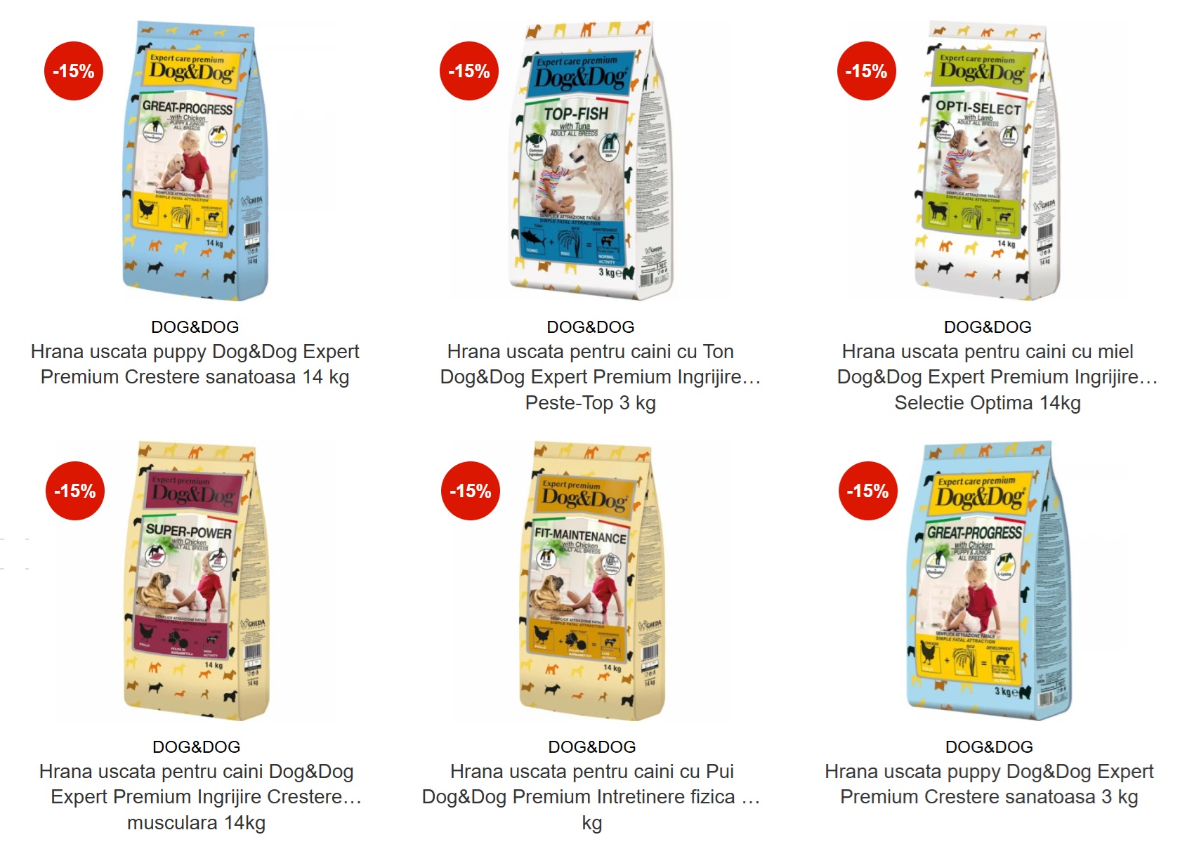
Everything you need to know about Dog & Dog – Dry Dog Food
Obesity in dogs and what should be done?
The dog needs to have a happy, balanced and healthy pet
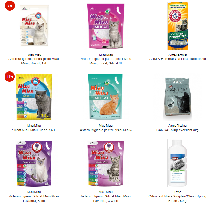
How do you choose and how do you use cat sand?
How do we help pets during winter holidays, full of strong noises and noises
The benefits of fish in the diet of our empty spaces
The joy of having a cat
Golden Retriever, an energetic and favorite breed of children